Retinoblastoma
Retinoblastoma

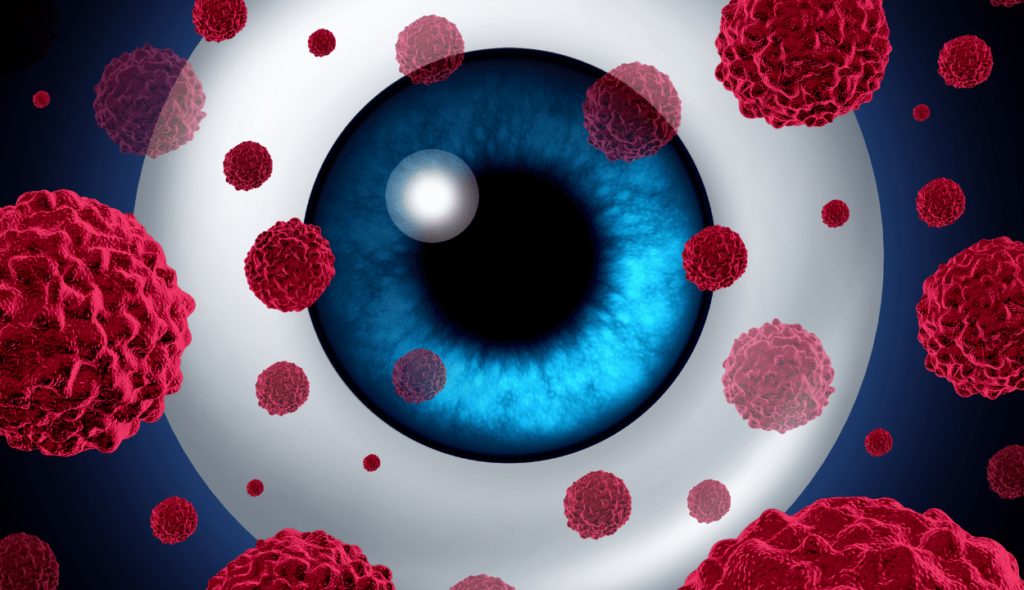
The retina, the delicate lining within the eye, is where retinoblastoma, an eye cancer, first appears. Although it seldom affects adults, retinoblastoma most frequently affects young children.
The nerve tissue that makes up your retina senses light as it enters your eye from the front. Your brain interprets the signals that the retina sends through the optic nerve as images.
Retinoblastoma, a rare kind of eye cancer, is the most prevalent type that affects children’s eyes. One or both eyes may develop retinoblastoma.
How does retinoblastoma develops?
Long before birth, the eyes begin to grow. Retinoblasts are cells that exist in the early stages of eye development and replicate to produce new retinal cells. These cells eventually cease proliferating and develop into mature retinal cells.
It happens very infrequently for this process to go awry. Some retinoblasts don’t mature; instead, they grow uncontrollably and develop into the malignancy retinoblastoma.
Retinoblastoma is caused by a complicated series of cellular events, but it virtually invariably begins with a change (mutation) in the RB1 gene. A mutation in the RB1 gene prevents it from functioning as it should, despite the fact that the normal RB1 gene aids in preventing uncontrolled cell growth. There are two main forms of retinoblastomas that can develop depending on where and when the RB1 gene is altered.
Types of retinoblastoma
Congenital (heritable) retinoblastoma
One in three children with retinoblastoma have a mutation in the RB1 gene that is congenital (existing at birth) and affects all body cells, including every retinal cell. A germline mutation is what this is.
Despite the fact that this is commonly referred to as “heritable” (or “hereditary”), the RB1 gene alteration is not passed down from parents in the majority of these children, and there is no family history of this cancer. The first time the gene changes in these kids is when they are still developing in the womb. The majority of kids who are born with this gene mutation do not get it from their parents.
Bilateral retinoblastoma, which affects both eyes in newborns with a mutation in the RB1 gene, frequently involves several tumours inside the eye (known as multifocal retinoblastoma).
Because all of the cells in the body have the changed RB1 gene, these children also have a higher risk of developing cancers in other parts of the body.
- A small number of children with this form of retinoblastoma will develop another tumor in the brain, usually in the pineal gland at the base of the brain (a pineoblastoma). This is also known as trilateral retinoblastoma.
- For survivors of hereditary retinoblastoma, the risk of developing other cancers later in life is also higher than average
Sporadic (non-heritable) retinoblastoma
The RB1 gene defect only manifests in one cell in one eye in around 2 out of 3 children with retinoblastoma. What triggers this shift is unknown. One tumour only appears in one eye in a child with sporadic (non-heritable) retinoblastoma. Compared to those who have the heritable form, this type of retinoblastoma is frequently discovered when the child is a little older.
The danger of additional cancers in children with this type of retinoblastoma is not as high as it is in those with congenital retinoblastoma.
Symptoms
Because retinoblastoma mostly affects infants and small children, symptoms aren’t common. Signs you may notice include:
- A white color in the center circle of the eye (pupil) when light is shone in the eye, such as when someone takes a flash photograph of the child
- Eyes that appear to be looking in different directions
- Poor vision
- Eye redness
- Eye swelling
Causes of retinoblastoma
When the retina’s nerve cells experience genetic abnormalities, retinoblastoma results. When healthy cells would die due to these mutations, the cells continue to grow and divide. A tumour develops from this growing clump of cells.
The eye and adjacent structures can be further invaded by retinoblastoma cells. The brain and spine are two other bodily parts where retinal blastoma can metastasis.
It is unclear what causes the genetic mutation that produces retinoblastoma in the majority of cases. However, a genetic mutation can be passed down to offspring from parents.
Diagnosis of retinoblastoma
Retinoblastomas are typically discovered when a youngster visits the doctor due to particular signs or symptoms.
A biopsy is required to make the diagnosis for the majority of cancer types. A sample of the tumour is removed during a biopsy and sent to a lab to be examined under a microscope. However, for two main reasons, biopsies are typically not performed to diagnose retinoblastoma.
It is difficult to take a biopsy sample from an eye tumour without endangering the eye and running the risk of cancer cells spreading outside the eye. Doctors with experience treating this condition can typically identify retinoblastoma accurately without doing a biopsy, and it is unlikely to be mistaken for other childhood eye conditions.
Physical examination
The doctor will check your child’s eyes and obtain a thorough medical history to learn more about your child’s symptoms if they show any retinoblastoma indications or symptoms. A family history of retinoblastoma or other tumours may also be brought up by the doctor. When determining whether more tests and exams are required, this can be crucial. Your family history can also help you determine whether additional family members might benefit from genetic testing and counselling if they have young children, potentially pass the retinoblastoma (RB1) gene change on to their offspring, or if they themselves have developed this cancer.
An ophthalmologist (a specialist who specialises in eye illnesses) will be referred to you if a retinoblastoma is suspected. The ophthalmologist will extensively inspect the eye to be more certain about the diagnosis. To examine the inside of the eye, the ophthalmologist will use specialised lighting and magnifying glasses. In order for the doctor to examine the child carefully and thoroughly, the youngster typically needs to be asleep (under general anaesthetic) during the procedure.
Imaging tests will be carried out to help confirm the diagnosis of retinoblastoma if it appears plausible based on the results of the eye exam. These tests will also help determine how far it may have progressed within the eye and maybe to other regions of the body. The final decision is typically made by an ocular oncologist, an ophthalmologist who specialises in treating eye cancers. The medical team treating the cancer should include this doctor as well.
Imaging tests
Imaging tests use x-rays, sound waves, magnetic fields, or radioactive substances to create pictures of the inside of the body. Imaging tests may be done for a number of reasons, including:
- To help tell if a tumor in the eye is likely to be a retinoblastoma
- To determine how large the tumor is and how far it has spread
- To help determine if treatment is working
Children thought to have retinoblastoma may have one or more of these tests.
Optical coherence tomography (OCT) is a similar type of test that uses light waves instead of sound waves to create very detailed images of the back of the eye.
MRI
Using radio waves and powerful magnets, an MRI scan produces precise images (instead of x-rays). Because they offer radiation-free, highly detailed images of the eye and its surroundings, MRI scans are frequently used to diagnose retinoblastomas. This examination is also excellent for examining the brain and spinal cord.
An MRI scan will typically be performed on retinoblastoma patients as part of their first evaluation. Many medical professionals continue to perform MRI scans of the brain in children who have bilateral retinoblastomas (tumours in both eyes) for years after treatment to check for pineal gland tumours (sometimes called trilateral retinoblastoma).
This test may require your child to lie inside a small tube, which is restricting and unpleasant. Long stretches of stillness are required for this test as well. To keep young children calm or even asleep during the test, medication may be administered to them.
CT Scan
A retinoblastoma tumor’s size and the extent of its dissemination within the eye and to surrounding tissues can both be determined using CT scans.
A CT or an MRI scan is typically required, but not both. Since CT scans employ x-rays, which could increase a child’s chance for developing other cancers in the future, most doctors choose to use MRI instead. When the diagnosis of retinoblastoma is unclear, a CT scan can reveal calcium deposits in the tumour much more clearly than an MRI.
Bone scan
If the retinoblastoma has progressed to the skull or other bones, a bone scan might assist reveal this. A bone scan is typically not necessary for children with retinoblastoma. Only when there is a good basis to believe that retinoblastoma may have spread outside of the eye is it typically used.
A small quantity of low-level radioactive material is injected into the blood for this test, where it goes to the bones. The radioactivity can be detected by a specialised camera, which then takes an image of the skeleton.
On the scan, “hot spots” represent regions of dynamic bone changes. These spots may indicate the presence of cancer, although the same pattern can also be caused by other bone illnesses. Other tests, such plain x-rays or MRI scans of the bone, may be required to assist distinguish between these.
Genetic testing
When a child is diagnosed with retinoblastoma, it’s critical to determine if the condition is heritable (congenital) or non-heritable.
It can be inferred that a child has heritable retinoblastoma if tumours are discovered in both eyes (bilateral retinoblastoma) (even if there is no family history of the disease). This indicates that every cell in them carries the RB1 mutant gene. The mutant RB1 gene may be present in all of the cells of some kids with retinoblastoma in just one eye.
To check for the RB1 gene alteration in cells beyond the eye, a blood test can be performed. This test can typically determine whether a child has retinoblastoma that is heritable.
It’s critical to understand which kind a child has since those who have heritable retinoblastoma are more susceptible to other cancers later in life and are more likely to acquire cancer if they receive radiation therapy. After treatment, these kids will require close monitoring. (View Following Treatment.) Additionally, each of their own offspring will have a 1 in 2 chance of inheriting the RB1 gene mutation from them.
Other family members, such as brothers or sisters, who may also possess the RB1 gene mutation, may be affected if a kid develops the heritable form of retinoblastoma. You can learn more about this risk and whether other children in the family should be tested for the mutation by meeting with a genetic counsellor.
Sometimes testing are unable to conclusively determine if a youngster received the RB1 gene mutation. The most secure course of action under these circumstances is to closely monitor the youngster (and other children in the family) for retinoblastoma through routine eye exams.
Biopsy
A biopsy, which involves taking a tissue sample from the tumour and examining it under a microscope, is required to diagnose the majority of malignancies. It is practically never done to identify retinoblastoma since attempting to biopsy a tumour at the back of the eye can frequently cause damage to the eye and may disseminate tumour cells. Instead, medical professionals base their diagnoses on imaging tests and eye exams like the ones mentioned above. This is why it’s crucial that retinoblastoma diagnoses are made by specialists.
Lumbar puncture
The optic nerve, which connects the eye to the brain, can occasionally develop retinoblastomas. This test frequently detects cancer cells in samples of cerebrospinal fluid (CSF), the fluid that surrounds the brain and spinal cord, if the cancer has migrated to the surface of the brain. A lumbar puncture is typically not necessary for children with retinoblastoma. It is mostly utilised when there is cause to suspect that retinoblastoma may have gone to the brain.
The youngster is typically given anaesthetic for this test so they will be sleepy and immobile during the process. This can assist guarantee a clean spinal tap. The lower back region over the spine is first made numb by the doctor. The fluid is then gently withdrawn and transported to the lab for analysis using a tiny, hollow needle that is inserted between the spine’s bones.
Bone marrow aspiration and biopsy
These 2 examinations could be carried out to determine whether the cancer has progressed to the bone marrow, the soft interior of some bones. Unless the retinoblastoma has progressed beyond the eye and medical professionals have reason to believe it may also have affected the bone marrow, these tests are typically not necessary.
Usually, the tests are completed simultaneously. In most cases, the samples are collected from the rear of the pelvic (hip) bone, although they can also come from other bones. The youngster is typically given anaesthetic so they are unconscious throughout the surgery.
A little amount of liquid bone marrow is aspirated with a syringe after being sucked out of the bone using a thin, hollow needle used for bone marrow aspiration.
Following the aspiration, a bone marrow biopsy is frequently performed. With a little larger needle that is driven down into the bone, a small bit of bone and marrow is extracted. Following completion of the biopsy, pressure is applied to the area to aid in stopping any bleeding.
Following that, a lab will examine the samples to check for cancerous cells.
Treatment of retinoblastoma
Surgery
For many retinoblastomas, especially those with smaller tumours, surgery is not necessary.
However, if a tumour has become huge when it is discovered, vision in the eye may have have been lost, with little chance of recovery. Enucleation, an operation to remove the entire eye and a portion of the optic nerve linked to it, is the typical course of treatment in this situation.
If other therapies intended to try to save the eye are unsuccessful in curing the cancer, enucleation may also be required.
The procedure is carried out with the youngster under general anaesthesia (in a deep sleep). The eyeball is typically replaced by an orbital implant during the same procedure. The implant is comprised of hydroxyapatite or silicone (a substance similar to bone). It should move in the same manner that the eye would have because it is connected to the muscles that moved the eye.
Most likely, your child will be allowed to leave the hospital that day or the following day.
You can take your child to an ocularist a few weeks later, and they will make a prosthetic eye for them. This is a thin shell that resembles a large contact lens and slips over the orbital implant and under the eyelids. It will be the same size and colour as the other eye. It will be quite difficult to distinguish it from the real eye once it is in place.
Enucleating both eyes would leave a person completely blind if retinoblastoma affected both eyes. This may be the best course of action to ensure that all cancer has been removed if neither eye has functional vision due to cancer-related damage previously done. However, doctors may suggest trying other forms of treatment first if there is a potential to preserve useful vision in one or both eyes.
Chemotherapy
Chemotherapy is a drug treatment that uses chemicals to kill cancer cells. In children with retinoblastoma, chemotherapy may help shrink a tumor so that another treatment, such as cryotherapy or laser therapy, may be used to treat the remaining cancer cells. This may improve the chances that your child won’t need surgery to remove the eye.
Types of chemotherapy used to treat retinoblastoma include:
- Chemotherapy that travels through the entire body. Chemotherapy drugs that are given through a blood vessel will travel throughout the body to kill cancer cells.
- Chemotherapy injected near the tumor. A specialized type of chemotherapy, known as intra-arterial chemotherapy, delivers the medicine directly to the tumor through a tiny tube (catheter) in an artery supplying blood to the eye. The doctor might put a tiny balloon in the artery to keep the medicine close to the tumor.
- Chemotherapy administered into the eye. Intravitreal chemotherapy involves injecting chemotherapy drugs directly into the eye.
Radiation therapy
Radiation therapy uses high-powered energy, such as X-rays and protons, to kill cancer cells. Types of radiation therapy used in treating retinoblastoma include:
Local radiation. During local radiation, also called plaque radiotherapy or brachytherapy, the treatment device is temporarily placed near the tumor.
Local radiation for retinoblastoma uses a small disk containing seeds of radioactive material. The disk is stitched in place and left for a few days while it slowly gives off radiation to the tumor.
Placing radiation near the tumor reduces the chance that treatment will affect healthy tissues outside the eye. This type of radiotherapy is typically used for tumors that don’t respond to chemotherapy.
External beam radiation. External beam radiation delivers high-powered beams to the tumor from a large machine outside of the body. As your child lies on a table, the machine moves around your child, delivering the radiation.
External beam radiation can cause side effects when radiation beams reach the delicate areas around the eye, such as the brain. For this reason, external beam radiation is typically reserved for children with advanced retinoblastoma.
Laser therapy (transpupillary thermotherapy)
During laser therapy, a heat laser is used to directly destroy the tumor cells.
Cold treatment (cryotherapy)
Cryotherapy uses extreme cold to kill cancer cells.
During cryotherapy, a very cold substance, such as liquid nitrogen, is placed in or near the cancer cells. Once the cells freeze, the cold substance is removed and the cells thaw. This process of freezing and thawing, repeated a few times in each cryotherapy session, causes the cancerous cells to die.
Take second opinion on retinoblastoma treatment
- Comments Closed
- June 27th, 2022


Latest Posts
- Targeting FGFR4 and CD276 with CAR T-cells demonstrates a strong antitumor impact against children rhabdomyosarcoma
- Disruption of CD5 on CAR T Cells Enhances the Effectiveness of Anti-Tumor Treatment
- The future of gene therapy: What to expect in the next decade?
- Unlocking the genetic code: The future of gene therapy for genetic disorders
- CRISPR and gene editing: Revolutionizing gene therapy
- Aids cancer (4)
- Anal cancer (8)
- Anemia (5)
- Appendix cancer (3)
- Basal cell carcinoma (1)
- Bile duct cancer (7)
- Bladder cancer (10)
- Blog (3)
- Blood cancer (56)
- Bone cancer (11)
- Bone marrow transplant (43)
- Brain Tumor (48)
- Breast Cancer (48)
- Cancer (787)
- Cancer surgery (234)
- Cancer treatment in South Korea (341)
- cancer treatment in Thailand (331)
- Cancer treatment in Turkey (329)
- Cancer treatment in USA (328)
- CAR NK-Cell therapy (12)
- CAR T-Cell therapy (95)
- Cervical cancer (41)
- Chemotherapy (36)
- Childhood cancer (2)
- Cholangiocarcinoma (3)
- Clinical trials (5)
- Colon cancer (95)
- Coronavirus (1)
- Cosmetic surgery (7)
- COVID19 (2)
- Doctor (37)
- Drugs (19)
- Endometrial cancer (9)
- Esophageal cancer (15)
- Eye cancer (9)
- Gall bladder cancer (3)
- Gastric cancer (22)
- Glioblastoma (1)
- Gynecological cancer (2)
- Head and neck cancer (20)
- Hematological Disorders (50)
- Hospital (47)
- Immunotherapy (25)
- Kidney cancer (10)
- Laryngeal cancer (1)
- Leukemia (44)
- Liver cancer (94)
- Lung cancer (65)
- Lymphoma (44)
- MDS (2)
- Medical tourism (71)
- Medical visa (11)
- Melanoma (8)
- Merkel cell carcinoma (1)
- Mesothelioma (4)
- Myeloma (22)
- Oral cancer (13)
- Ovarian Cancer (13)
- Pancreatic cancer (39)
- Penile cancer (1)
- Procedure (19)
- Prostrate cancer (10)
- Proton therapy (26)
- Radiotherapy (35)
- Rectal cancer (57)
- Sarcoma (11)
- Skin Cancer (13)
- Spine surgery (8)
- Stomach cancer (40)
- Surgery (54)
- Systemic mastocytosis (1)
- T Cell immunotherapy (2)
- T-Cell therapy (7)
- Testicular cancer (5)
- Thoracic surgery (2)
- Throat cancer (6)
- Thyroid Cancer (14)
- Treatment (746)
- Treatment in China (646)
- Treatment in India (684)
- Treatment in Israel (586)
- Treatment in Malaysia (360)
- Treatment in Singapore (255)
- Treatment in South Korea (238)
- Treatment in Thailand (233)
- Treatment in Turkey (233)
- Uncategorized (39)
- Urethral cancer (9)
- Urosurgery (14)
- Uterine cancer (3)
- Vaginal cancer (6)
- Vascular cancer (5)
- Vulvar cancer (1)


Privacy Overview