Gallbladder cancer
About Disease

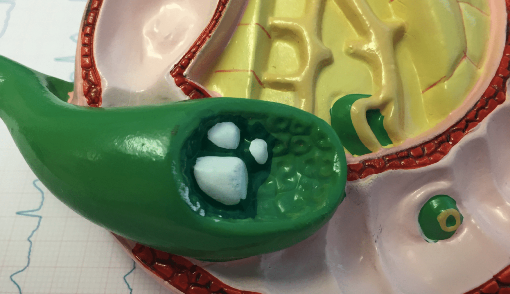
A malignant cell growth that starts in the gallbladder is known as gallbladder cancer. On the right side of your belly, beneath your liver, is a little, pear-shaped organ called the gallbladder. Bile, a digestive fluid created by your liver, is kept in the gallbladder.
Cancer of the gallbladder is rare. The likelihood of a cure is relatively high when gallbladder cancer is found in its earliest stages. However, the majority of gallbladder cancers are found in the late stages, when the prognosis is frequently very dismal.
Because gallbladder cancer frequently exhibits no particular signs or symptoms, it might not be identified until it has progressed. Gallbladder cancer is also more likely to spread undetected due to the gallbladder’s relative secrecy.
Malignant (cancer) cells are discovered in the tissues of the gallbladder in the rare condition known as gallbladder cancer. The gallbladder is a pear-shaped organ that is located in the upper belly next to the liver. Bile, a substance produced by the liver to break down fat, is kept in the gallbladder. The common bile duct, which links the gallbladder, liver, and first portion of the small intestine, allows bile from the gallbladder to be released when food is digested in the stomach and intestines.
Stages of gallbladder cancer
Stage 0 (Carcinoma in Situ)
In stage 0, abnormal cells are found in the mucosa (innermost layer) of the gallbladder wall. These abnormal cells may become cancer and spread into nearby normal tissue. Stage 0 is also called carcinoma in situ.
Stage I
In stage I, cancer has formed in the mucosa (innermost layer) of the gallbladder wall and may have spread to the muscle layer of the gallbladder wall.
Stage II
Stage II is divided into stages IIA and IIB, depending on where the cancer has spread in the gallbladder.
- In stage IIA, cancer has spread through the muscle layer to the connective tissue layer of the gallbladder wall on the side of the gallbladder that is not near the liver.
- In stage IIB, cancer has spread through the muscle layer to the connective tissue layer of the gallbladder wall on the same side as the liver. Cancer has not spread to the liver.
Stage III
Stage III is divided into stages IIIA and IIIB, depending on where the cancer has spread.
- In stage IIIA, cancer has spread through the connective tissue layer of the gallbladder wall and one or more of the following is true:
- Cancer has spread to the serosa (layer of tissue that covers the gallbladder).
- Cancer has spread to the liver.
- Cancer has spread to one nearby organ or structure (such as the stomach, small intestine, colon, pancreas, or the bile ducts outside the liver).
- In stage IIIB, cancer has formed in the mucosa (innermost layer) of the gallbladder wall and may have spread to the muscle, connective tissue, or serosa (layer of tissue that covers the gallbladder) and may have also spread to the liver or to one nearby organ or structure (such as the stomach, small intestine, colon, pancreas, or the bile ducts outside the liver). Cancer has spread to one to three nearby lymph nodes.
Stage IV
Stage IV is divided into stages IVA and IVB.
- In stage IVA, cancer has spread to the portal vein or hepatic artery or to two or more organs or structures apart from the liver. Cancer may have spread to one to three nearby lymph nodes.
- In stage IVB, cancer may have spread to nearby organs or structures. Cancer has spread:
- to four or more nearby lymph nodes; or
- to other parts of the body, such as the peritoneum and liver.
Overview
Causes
The exact cause of gallbladder cancer is unknown. Doctors are aware that the development of DNA abnormalities in healthy gallbladder cells leads to the development of gallbladder cancer. The instructions that inform a cell what to do are encoded in its DNA.
The modifications instruct the cells to proliferate unchecked and to live longer than other cells would otherwise. The collecting cells create a tumor, which has the potential to move outside of the gallbladder and to other parts of the body.
Factors that can increase the risk of gallbladder cancer include:
- Your sex: Gallbladder cancer is more common in women.
- Your age: Your risk of gallbladder cancer increases as you age.
- A history of gallstones: Gallbladder cancer is most common in people who have gallstones or have had gallstones in the past. Larger gallstones may carry a larger risk. Still, gallstones are very common, and even in people with this condition, gallbladder cancer is very rare.
- Other gallbladder diseases and conditions: Other gallbladder conditions that can increase the risk of gallbladder cancer include polyps, chronic inflammation, and infection.
- Inflammation of the bile ducts: Primary sclerosing cholangitis, which causes inflammation of the ducts that drain bile from the gallbladder and liver, increases the risk of gallbladder cancer.
The glandular cells that line the inner surface of the gallbladder are where the majority of gallbladder cancers start. Adenocarcinoma is the name for gallbladder cancer that develops in these cells. When cancer cells are inspected under a microscope, they take on this description.
Symptoms
- There are no signs or symptoms in the early stages of gallbladder cancer.
- The symptoms of gallbladder cancer, when present, are like the symptoms of many other illnesses.
- The gallbladder is hidden behind the liver.
Gallbladder cancer is sometimes found when the gallbladder is removed for other reasons. Patients with gallstones rarely develop gallbladder cancer.
These and other signs and symptoms may be caused by gallbladder cancer or by other conditions. Check with your doctor if you have any of the following:
- Jaundice (yellowing of the skin and whites of the eyes).
- Pain above the stomach
- Fever
- Nausea and vomiting
- Bloating
- Lumps in the abdomen
Diagnosis
Procedures that make pictures of the gallbladder and the area around it help diagnose gallbladder cancer and show how far the cancer has spread. The process used to find out if cancer cells have spread within and around the gallbladder is called staging.
To plan treatment, it is important to know if the gallbladder cancer can be removed by surgery. Tests and procedures to detect, diagnose, and stage gallbladder cancer are usually done at the same time. The following tests and procedures may be used:
- Physical exam and health history: An exam of the body to check general signs of health, including checking for signs of disease, such as lumps or anything else that seems unusual. A history of the patient’s health habits and past illnesses and treatments will also be taken.
- Liver function tests: A procedure in which a blood sample is checked to measure the amounts of certain substances released into the blood by the liver. A higher-than-normal amount of a substance can be a sign of liver disease that may be caused by gallbladder cancer.
- Blood chemistry studies: A procedure in which a blood sample is checked to measure the amounts of certain substances released into the blood by organs and tissues in the body. An unusual (higher or lower than normal) amount of a substance can be a sign of disease.
- CT scan (CAT scan): A procedure that makes a series of detailed pictures of areas inside the body, such as the chest, abdomen, and pelvis, taken from different angles. The pictures are made by a computer linked to an x-ray machine. A dye may be injected into a vein or swallowed to help the organs or tissues show up more clearly. This procedure is also called computed tomography, computerized tomography, or computerized axial tomography.
- Ultrasound exam: A procedure in which high-energy sound waves (ultrasound) are bounced off internal tissues or organs and make echoes. The echoes form a picture of body tissues called a sonogram. An abdominal ultrasound is done to diagnose gallbladder cancer.
- PTC (percutaneous transhepatic cholangiography): A procedure used to x-ray the liver and bile ducts. A thin needle is inserted through the skin below the ribs and into the liver. Dye is injected into the liver or bile ducts, and an x-ray is taken. If a blockage is found, a thin, flexible tube called a stent is sometimes left in the liver to drain bile into the small intestine or a collection bag outside the body.
- ERCP (endoscopic retrograde cholangiopancreatography): A procedure used to x-ray the ducts (tubes) that carry bile from the liver to the gallbladder and from the gallbladder to the small intestine. Sometimes gallbladder cancer causes these ducts to narrow and block or slow the flow of bile, causing jaundice. An endoscope (a thin, lighted tube) is passed through the mouth, esophagus, and stomach into the first part of the small intestine. A catheter (a smaller tube) is then inserted through the endoscope into the bile ducts. A dye is injected through the catheter into the ducts and an x-ray is taken. If the ducts are blocked by a tumor, a fine tube may be inserted into the duct to unblock it. This tube (or stent) may be left in place to keep the duct open. Tissue samples may also be taken.
- MRI (magnetic resonance imaging) with gadolinium: A procedure that uses a magnet, radio waves, and a computer to make a series of detailed pictures of areas inside the body. A substance called gadolinium is injected into a vein. The gadolinium collects around the cancer cells so they show up brighter in the picture. This procedure is also called nuclear magnetic resonance imaging (NMRI).
- Endoscopic ultrasound (EUS): A procedure in which an endoscope is inserted into the body, usually through the mouth or rectum. An endoscope is a thin, tube-like instrument with a light and a lens for viewing. A probe at the end of the endoscope is used to bounce high-energy sound waves (ultrasound) off internal tissues or organs and make echoes. The echoes form a picture of body tissues called a sonogram. This procedure is also called endosonography.
- Laparoscopy: A surgical procedure to look at the organs inside the abdomen to check for signs of disease. Small incisions (cuts) are made in the wall of the abdomen, and a laparoscope (a thin, lighted tube) is inserted into one of the incisions. Other instruments may be inserted through the same or other incisions to perform procedures such as removing organs or taking tissue samples for biopsy. The laparoscopy helps to find out if the cancer is within the gallbladder only or has spread to nearby tissues and if it can be removed by surgery.
- Biopsy: The removal of cells or tissues so they can be viewed under a microscope by a pathologist to check for signs of cancer. The biopsy may be done after surgery to remove the tumor. If the tumor clearly cannot be removed by surgery, the biopsy may be done using a fine needle to remove cells from the tumor.
Treatment and Management
- There are different types of treatment for patients with gallbladder cancer.
- Three types of standard treatment are used:
- Surgery
- Radiation therapy
- Chemotherapy
- New types of treatment are being tested in clinical trials.
- Radiation sensitizers
- Targeted therapy
- Immunotherapy