
Curious about how a medical technique is saving the lives of multiple myeloma patients? Read our blog to learn more about this life-saving technique! Let’s turn the page on cancer together. Your story of hope starts here.
Hello there, everyone! Today, we’re going to talk about a special kind of cancer disease called multiple myeloma and how doctors use diagnostic imaging in multiple myeloma to understand and fight it.
Multiple myeloma is a type of blood cancer that demands a comprehensive approach for effective diagnosis and treatment. As you continue reading this article further, you will understand the significance of diagnostic imaging, and also discover how these powerful multiple myeloma treatment in India enable healthcare providers to make informed decisions for effective and personalized treatment.
So, if you or your loved ones are facing this challenging situation, you will find this blog helpful. Ready for an informative journey where you will learn how pictures save the lives of multiple myeloma patients? Let’s dive in!
What Is Multiple Myeloma?
Multiple myeloma is a kind of cancer that begins in plasma cells, which are immune system components present in bone marrow. These cells, responsible for producing antibodies, undergo abnormal multiplication, leading to the overcrowding of healthy cells. This disturbance weakens bones and impairs immune function. Diagnosis of multiple myeloma in the early stage is very crucial for effective treatment. Understanding the characteristics and behavior of this cancer type allows oncologists to take measures to combat its impact on the body, promoting a more targeted and successful approach. Right now, reputed cancer hospitals like TATA Cancer Research Center have already started providing CAR T Cell therapy treatment in India to prevent the progression of various types of blood cancer. Moreover, the cost of CAR T cell therapy in India makes it a better option for advanced cancer treatment.
If you want to know more about this revolutionary therapy, read this blog:
The Role Of Immunotherapy In Lymphoma Treatment – CancerFax

Importance Of Diagnostic Imaging In Multiple Myeloma
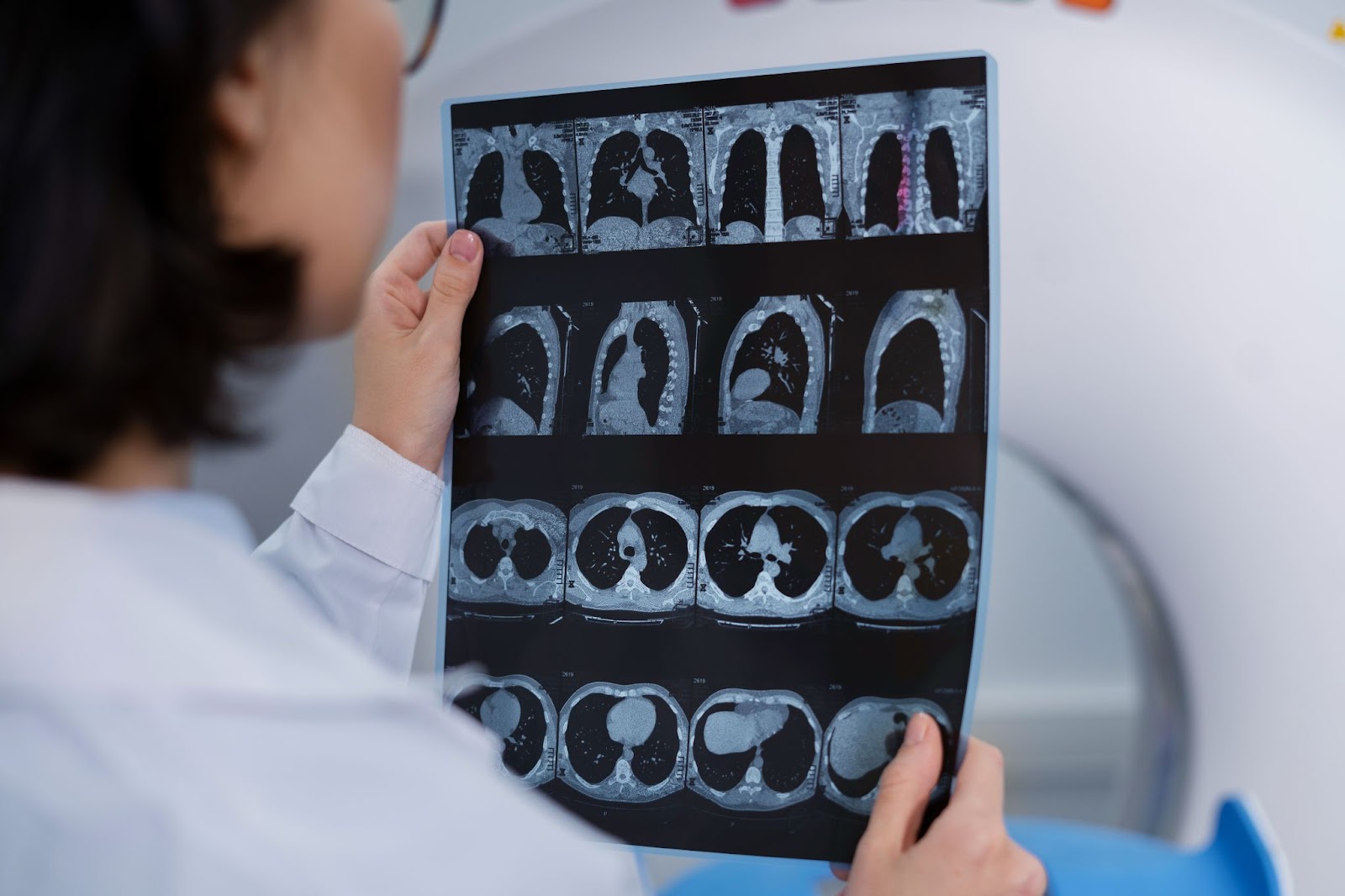
Diagnostic imaging plays an important role in the comprehensive management of multiple myeloma, providing important insights into the disease’s complexities. Various imaging techniques, such as X-rays, CT scans, MRIs, and PET scans, collectively contribute to a comprehensive overview of the affected areas within the body. One of the key benefits of diagnostic imaging is its ability to aid in early detection. These approaches operate as an alert eye, detecting even small abnormalities or signs of myeloma at an earlier and more curable stage. Beyond early detection, the true power of diagnostic imaging lies in its capacity to inform personalized treatment plans. By clearly visualizing the location, extent, and features of the myeloma, healthcare providers can tailor therapies to the individual needs of each patient.
Must Read: Immunotherapy Can Help You Win The Battle Against Multiple Myeloma!
Types Of Diagnostic Imaging In Multiple Myeloma Treatment
Explore the diagnostic imaging landscape in multiple myeloma treatment, from the precision of X-rays and CT scans to the detailed insights provided by MRI and PET scans. Learn how these imaging techniques guide personalized strategies for effective multiple myeloma treatment in India.
X-rays
X-rays, the pioneers of diagnostic imaging, play an essential role in the treatment of multiple myeloma. These images reveal the interior structure of bones, allowing healthcare practitioners to detect irregularities or lesions that are symptomatic of myeloma. While X-rays are a traditional diagnostic technique, their capacity to capture bone features remains invaluable. They are especially useful in detecting bone deterioration, fractures, or the presence of lytic lesions, which provides valuable information for staging and treatment planning.

Computed Tomography Scans
Computed tomography (CT) scans are a powerful method for the detection of multiple myeloma. Low-dose whole-body CT scans, as opposed to standard skeletal surveys, are proposed as a more sensitive option, particularly for detecting lesions in difficult places such as the ribs, pelvis, or spine. These scans are capable of identifying even the smallest lytic bone abnormalities that regular X-rays may miss, and they can even predict the likelihood of fractures. While CT scans are effective for detecting complications and providing thorough information, they have limitations, especially in detecting bone marrow tumors and widespread disorders. Still, their ability to uncover hidden details makes them an invaluable tool in the fight against multiple myeloma.
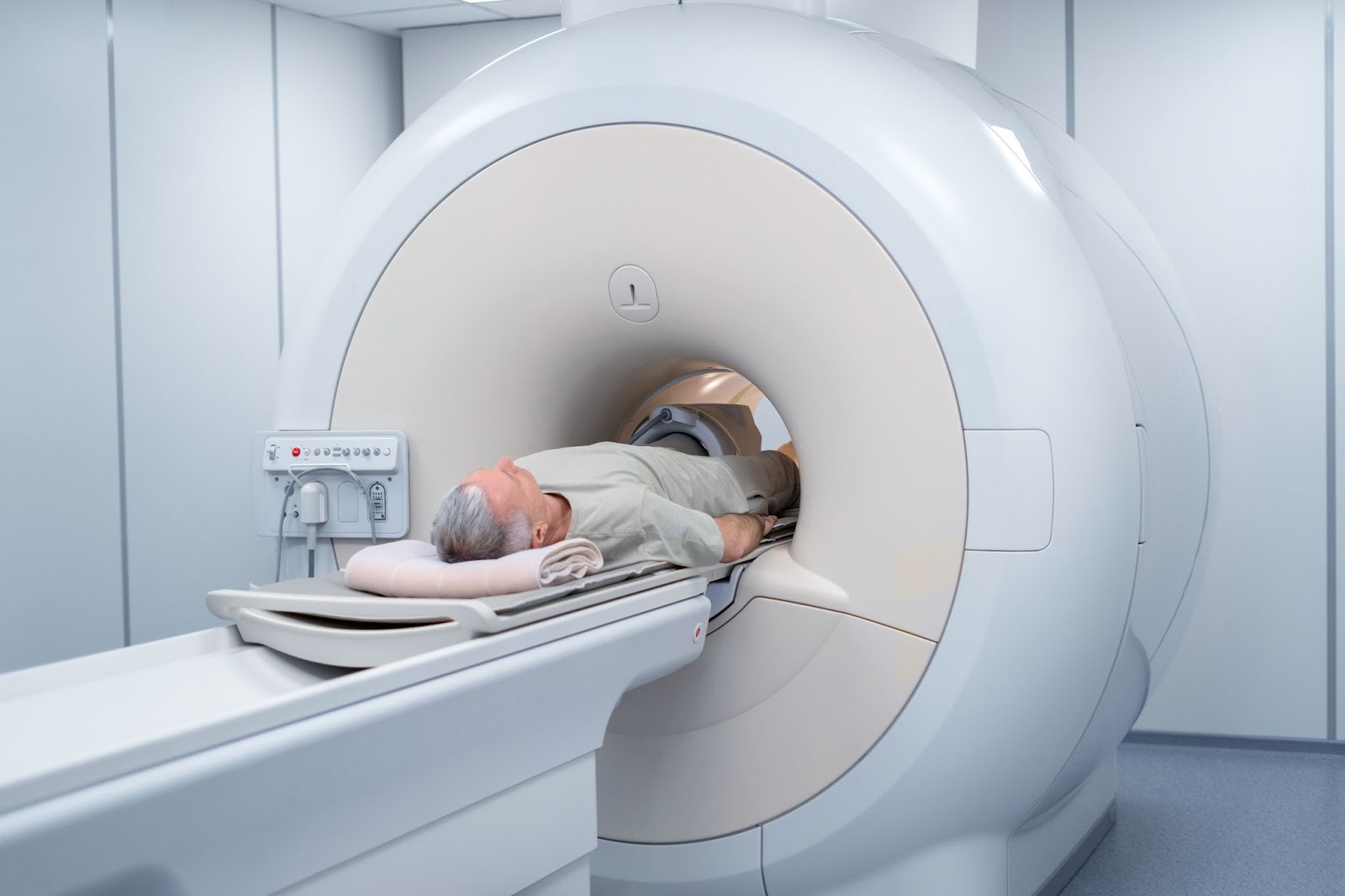
Magnetic Resonance Imaging
Magnetic Resonance Imaging (MRI) plays an important role in the staging process for people with multiple myeloma. Its higher sensitivity in detecting lesions, compared to traditional skeletal surveys and other imaging methods, makes it an essential tool in the diagnostic toolkit. MRI offers an unparalleled view of the bone marrow, enabling early detection of marrow infiltration even before myeloma-related bone damage occurs. When it comes to complete imaging, Whole-body MRI (WB-MRI) takes the lead, outperforming conventional MRI of specific areas such as the spine and pelvis. This innovative imaging technology is crucial in giving specific insights that guide healthcare practitioners in the accurate evaluation and management of multiple myeloma.
Positron Emission Tomography Scans
Positron Emission Tomography (PET) scans, particularly those utilizing fluorodeoxyglucose (FDG) as a tracer, have become a standard method in the evaluation of multiple myeloma patients. The importance of FDG PET imaging lies in its ability to offer a comprehensive view of the entire body, helping in the assessment of tumors and distinguishing between metabolically active and inactive lesions. FDG is taken by hyperactive, malignant plasma cells as a substitute for glucose. When PET scans are paired with CT scans, they constitute a dynamic duo that provides not only functional information regarding metabolic activity but also precise anatomical localization of abnormalities. This data fusion improves diagnosis accuracy, allowing healthcare practitioners to personalize treatment strategies more effectively in the fight against multiple myeloma.
Gain Insights On : Patient Selection For CAR T Therapy: A COMPLETE GUIDE
Benefits Of Diagnostic Imaging In Multiple Myeloma
- Imaging techniques help in the early detection of myeloma-related abnormalities and allow for faster intervention and improved treatment outcomes.
- Imaging is essential in staging because it provides a comprehensive view of the extent and severity of the disease.
- Different imaging techniques help to understand the location, size, and features of myeloma lesions.
- Diagnostic imaging helps in determining the treatment response in the patient’s body.
- Imaging techniques can detect complications associated with multiple myelomas, such as fractures or other skeletal issues.
- It minimizes potential side effects and improves the overall quality of life for people with multiple myeloma.
Note: This blog is intended to provide general information about the crucial role of diagnostic imaging in the context of multiple myeloma. It is important to note that the content offered here is not a substitute for professional medical advice, diagnosis, or treatment. Please consult a healthcare provider to know what the best solution in your case is.
Final Thoughts:
That brings us to the end of this informative article on diagnostic imaging in multiple myeloma. Who knew images could be so powerful? Now you have the knowledge to get the best multiple myeloma treatment in India. However, always consider the opinion of your oncologist to win the fight against this serious health condition. We wish you a speedy recovery and a brighter tomorrow!

Dr. Nishant Mittal is a highly accomplished researcher with over 13 years of experience in the fields of cardiovascular biology and cancer research. His career is marked by significant contributions to stem cell biology, developmental biology, and innovative research techniques.
Research Highlights
Dr. Mittal's research has focused on several key areas:
1) Cardiovascular Development and Regeneration: He studied coronary vessel development and regeneration using zebrafish models1.
2) Cancer Biology: At Dartmouth College, he developed zebrafish models for studying tumor heterogeneity and clonal evolution in pancreatic cancer.
3) Developmental Biology: His doctoral work at Keio University involved identifying and characterizing medaka fish mutants with cardiovascular defects.
4) Stem Cell Research: He investigated the effects of folic acid on mouse embryonic stem cells and worked on cryopreservation techniques for hematopoietic stem cells.
Publications and Presentations
Dr. Mittal has authored several peer-reviewed publications in reputable journals such as Scientific Reports, Cardiovascular Research, and Disease Models & Mechanisms1. He has also presented his research at numerous international conferences, including the Stanford-Weill Cornell Cardiovascular Research Symposium and the Weinstein Cardiovascular Development Conference.
In summary, Dr. Nishant Mittal is a dedicated and accomplished researcher with a strong track record in cardiovascular and cancer biology, demonstrating expertise in various model systems and a commitment to advancing scientific knowledge through innovative research approaches.

