A PET scan is a ray of hope in the fight against cancer. If your doctor suspects a case of cancer, they might use this imaging method for an accurate diagnosis. Read our informative guide to learn all about this revolutionary technology shaping a hopeful future for cancer patients worldwide.
Positron Emission Tomography, or PET CT scans, has become a ground-breaking technology in the field of medical diagnostics, revolutionizing our understanding and treatment of cancer. This modern imaging technique combines Positron Emission Tomography (PET) and Computed Tomography (CT) to provide a complete view of the body’s internal structures and functions.
In this article, let’s try to understand the importance of PET CT scans and how they’re becoming a lifeline for cancer patients throughout the world, providing new hope and accuracy in the diagnosis and treatment of this challenging disease.
What Is A Pet CT Scan?
A PET CT scan is a medical miracle that combines Computed Tomography and Positron Emission Tomography. It is essential for diagnostic purposes, especially when fighting cancer.
During this procedure, a tiny amount of radioactive material, known as a radiotracer, is introduced into the body. This radiotracer acts like a detective, seeking out areas with unusual activity, such as tumors or inflammation.
One common radiotracer, F-18 fluorodeoxyglucose (FDG), mimics glucose and is especially effective at highlighting rapidly dividing cancer cells. The emitted gamma rays from the radiotracer are then captured by a special camera, producing detailed images
During this process, a specific dye containing radioactive tracer is used. Depending on the area of the body being examined, these tracers are swallowed, inhaled, or injected into an arm vein. Tracers are absorbed by certain organs and tissues after they have been injected.
The tracers are more likely to accumulate in regions with greater chemical activity, which is important because certain tissues and diseases have higher chemical activity than others.
As a result, the PET scan shows these regions of heightened activity as bright spots, which helps medical professionals identify possible areas of concern.
What makes PET CT scans even more powerful is their ability to collaborate with other imaging techniques such as CT or MRI, resulting in a holistic image that helps doctors establish precise diagnoses and treatment plans.
This non-invasive procedure not only allows for early disease detection but also provides crucial insights into the effectiveness of ongoing treatments.
Why Do Doctors Prescribe PET Scan Imaging?
Doctors may recommend PET imaging to get a better understanding of how your body works at the cellular level. These scans provide a complete picture of complex systemic disorders by looking at parameters such as blood flow, oxygen intake, and organ and tissue metabolism.
One major benefit is their ability to reveal cancer cells due to their higher metabolic rate, showing up as bright spots on the scan. PET scans are not only helpful in detecting cancer but also serve various purposes such as checking if cancer has spread, monitoring the effectiveness of cancer treatment like chemotherapy, and keeping an eye out for cancer recurrence.
However, these scans must be carefully interpreted by a specialist, as noncancerous illnesses can sometimes mimic cancer appearances on the scan, and certain solid tumors may not be visible.
How Does PET CT Scan Work?
During a PET-CT scan, a small injection of radioactive sugar, called fluorodeoxyglucose-18 (FDG-18), is given to you. This sugar is absorbed by your body’s cells, and areas using more energy, like cancer cells, absorb it more.
The PET scan then reveals where the radioactive sugar is in your body. Additionally, a CT scan involves taking X-rays from different angles, and a dye may be given before the X-rays to enhance visibility.
Combining the PET and CT pictures on a computer generates a full 3-D report for your doctor. This report indicates any irregularities, making it easier for the doctor to detect conditions such as tumors, which have higher energy consumption than normal cells.
How To Prepare For A Pet CT Scan?
Clothing And Accessories
Dress appropriately, either in a gown or according to the instructions provided.
Inform About Pregnancy And Breastfeeding
If you are pregnant or breastfeeding, notify your doctor and the CT scanner.
Medication And Health Disclosure
Disclose all medications, vitamins, and herbal supplements you take.
Inform about allergies, recent illnesses, or other health issues.
Special Instructions For Diabetes
Patients with diabetes will be given specific preparation guidelines.
Breastfeeding Considerations
If you are breastfeeding, get advice and consider pumping your milk a few hour before the scan.
Metal Items And Accessories
Leave metal items, such as jewelry and eyeglasses, at home.
Remove hearing aids and dental procedures as needed.
Fasting Before PET/CT Scan
Follow fasting instructions before a full-body PET/CT scan.
Avoid sugary or calorie-containing liquids; drink water as directed.
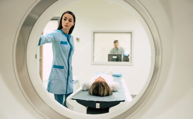
How Is Positron Emission Tomography Scan Performed?
Before a Positron Emission Tomography (PET) scan, you’ll receive tracers through a vein in your arm, a solution you drink, or inhale as a gas. After taking the tracers, you wait for about an hour to let your body absorb them, the time depending on the scanned area.
During this time, it’s good to limit movement and stay warm. For the actual scan, which takes 30 to 45 minutes, you lie on a narrow table attached to a PET machine shaped like a giant “O.” The table goes gently into the machine for scanning. If multiple tests are needed, it might take up to 3 hours.
During the scan, you must lie still and follow the technician’s instructions, which include holding your breath for a few seconds. You’ll hear buzzing and clicking sounds. Once all images are recorded, you slide out of the machine, and the test is complete.
Is PET CT Scan Safe For Cancer Diagnosis?
Yes, a PET CT scan is generally safe and helps diagnose cancer effectively. Although it uses radioactive tracers, the exposure to harmful radiation is minimal, and the amount in the tracer is small, posing low risks to your body.
These tracers meet strict safety and performance standards set by the Food and Drug Administration (FDA). It’s always a good idea to discuss any concerns with your doctor, but the risks of the test are minimal compared to the valuable results it provides for diagnosing serious medical conditions.
Key Benefits Of Positron Emission Tomography And Computed Tomography
1. PET-CT scans produce precise images, allowing clinicians to detect cancer and its exact location in the body.
2. These scans help to determine the stage of cancer, including how far it has progressed and whether it has spread to other places.
3. PET-CT helps doctors evaluate the effectiveness of cancer treatments, such as chemotherapy, by observing changes in tumor activity.
4. After therapy, these scans can detect if cancer has reappeared, allowing for earlier action.
5. PET-CT combines PET’s metabolic information with CT’s detailed anatomical images, providing a more comprehensive view for improved diagnostic accuracy.
6. PET-CT is valuable in spotting cancer at an early stage, enhancing the chances of successful treatment and improved outcomes.
How much Does A PET CT Scan cost?
To know the cost of PET CT scans in different countries, please visit here and select PET Scan in treatment followed by the name of the country.
To Sum Up:
In a nutshell, PET CT scans are making a huge difference for people fighting cancer worldwide. They help doctors find and understand cancer better, making treatments more effective.
From spotting cancer early to creating personalized plans, these scans are changing the way we fight against the disease. By giving us a clearer picture of what’s happening inside the body, PET CT scans are not just improving lives but also offering new hope in the global battle against cancer.